
Synthesis and characterization of ultrasmall AuPd nanozymes
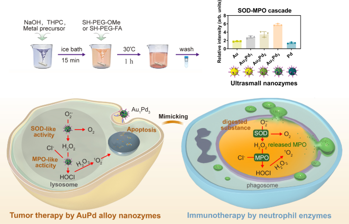
Ultrasmall AuPd alloy nanozymes were prepared through a modified method based on P(CH2OH)4Cl (tetrakis (hydroxymethyl) phosphonium chloride, THPC)19,20 (Fig. 1). The alloy ratio was regulated by adjusting the proportions of metal precursors. The surface of the ultrasmall AuPd alloy nanozymes was coated with SH-PEG-OMe through strong coordination between metal and thiol (SH) to maintain stability and dispersibility, as well as reduce non-specific adsorption of proteins21,22. High-resolution transmission electron microscopy (HRTEM) images demonstrated the spherical morphology and uniform distribution of the synthesized five nanozymes, all exhibiting a diameter range of 2-3ânm. (Fig. 2a). The Pd content in the five nanozymes was determined by an inductively coupled plasma optical emission spectrometer (ICP-OES). The results showed that the five nanozymes contained increasing amounts of Pd, indicating that we successfully obtain five nanozymes with different alloy ratios (Fig. 2b). These nanozymes were labeled as Au, Au3Pd1, Au2Pd2, Au1Pd3, and Pd, corresponding to their respective alloy compositions. The coverage of SH-PEG-OMe was confirmed by Fourier transform infrared (FTIR), which showed strong bands at 2890âcmâ1 and 1112âcmâ1 related to the C-H and C-O stretching vibrations of PEG main chain, respectively (Fig. 2c)23. The interaction between SH-PEG-OMe and AuPd alloy nanozymes through the Au-S bond or Pd-S bond was verified by the appearance of peaks at about 300âcmâ1 in the Raman spectra (Fig. 2d)24,25. The structures of ultrasmall AuPd alloy nanozymes were determined by powder X-ray diffraction (XRD) (Fig. 2e). The XRD patterns of Au and Pd nanozymes can be indexed to the face-centered cubic Au and Pd, and the peaks of Au3Pd1, Au2Pd2 and Au1Pd3 alloy nanozymes showed a shift from Au (PDF#04-0784) to Pd (PDF#46-1043), demonstrating the formation of alloy structures. X-ray photoelectron spectroscopy (XPS) was used to analyze the effect of alloying on the chemical states of AuPd alloy nanozymes. It can be seen from the XPS spectra that all five nanozymes contained related metal elements and C, O elements that constitute SH-PEG-OMe. Additionally, Na was present in the nanozymes due to the addition of NaOH during the synthesis process (Supplementary Fig. 1). The valence states of Au in Au nanozymes encompassed Au0, Au1+, and Au3+, as illustrated in Fig. 2f. Similarly, Pd nanozymes exhibited the valence states of Pd0 and Pd2+. These results suggest that the surface of Au nanozymes undergo the adsorption of Au[Clx(OH)4âx]â species, while Pd nanozymes experience the adsorption of Pd[Clx(OH)y]2âxây species26,27. Pd in AuPd alloy nanozymes would exhibit a certain degree of electropositivity because of the higher electronegativity of Au28,29. As the proportion of Pd on the surface increased from Au to Au3Pd1 nanozymes, the percentage of Au3+ increased because the positively charged Pd adsorbed more Au[Clx(OH)4-x]â, in which Au refers to Au3+. The coverage of SH-PEG-OMe made the five nanozymes stable in solution, with average hydrodynamic diameters of 3.8â5.3ânm (Fig. 2g). Their ultrasmall size (less than 6ânm) gives them the potential for renal clearance30. Above results show that we successfully synthesized nanozymes with ultrasmall size and different alloy ratios.

a HRTEM images, size distribution (nâ=â100 nanoparticles) and schematic illustration of five ultrasmall nanozymes. Scale bars = 5ânm. b The Pd content in five AuPd alloy nanozymes determined by ICP-OES (nâ=â3 independent experiments). c FTIR spectra of five AuPd nanozymes and SH-PEG-OMe. d Raman spectra of five AuPd nanozymes. e XRD patterns and (f) XPS spectra of five AuPd nanozymes. g Hydrodynamic diameter distribution of five ultrasmall nanozymes. All data are presented as meanâ±âSTD. Source data are provided as a Source Data file.
Imitation of the cascade catalysis of neutrophils by AuPd nanozymes
It has been reported that alloying can finely tune the catalytic activity of metals18. Thus, we investigated the enzyme-like activities of five AuPd nanozymes and explored the relationship between the activity and alloy ratio. Interestingly, we found that these nanozymes exhibited both SOD- and MPO-like activities simultaneously, and their activities were closely related to the alloy ratio. The SOD-like activity was determined by a SOD assay kit, and normalized by the molar concentration to compare the difference between nanozymes with different degrees of alloying. As shown in Fig. 3a, the SOD-like activity of either Au or Pd nanozymes was low, and alloying could increase their activity. Au1Pd3 alloy nanozymes exhibited the highest SOD-like activity, with a specific activity of about 7300 U mgâ1, which is much higher than that of natural SOD31. It is supposed that as the Pd content in the alloy increased, the SOD-like activity of AuPd nanozymes should exhibit a pattern of initially increasing and then decreasing, with the corresponding peak ratio being the optimal ratio. In order to determine the alloy ratio of nanozymes with the optimal SOD-like activity, we further prepared the Au1.3Pd8.7 alloy nanozymes, with the Pd content in the alloy determined to be 87% by the ICP-OES analysis. As shown in Supplementary Fig. S2, the Au1.3Pd8.7 nanozymes consisted of uniformly distributed small particles with a hydrated particle size of 3.8ânm, which was consistent with the other five nanozymes. The results of the SOD activity assay demonstrated that the Au1.3Pd8.7 alloy nanozymes possessed lower activity than the Au1Pd3 alloy nanozymes, but higher activity than the Pd nanozymes. These results suggest that the alloy nanozymes with an Au:Pd ratio of 1:3 exhibit the optimal SOD-like activity. The MPO-like activities of AuPd nanozymes were detected by monitoring the conversion of monochlorodimedon (MCD) to dichlorodimedon32, using the change in absorbance of MCD at 290ânm as an indicator (Supplementary Fig. 3). The statistical results showed that the MPO-like activities of AuPd nanozymes increased with the increase of the proportion of Pd (Fig. 3b). The high SOD- and MPO-like activities of the AuPd alloy nanozymes also benefit from their ultrasmall size, as we found that the ultrasmall-sized AuPd alloy nanozymes exhibited higher enzymatic activities compared to the large-sized AuPd alloy nanozymes (Supplementary Fig. 4), which may be related to their higher specific surface area, potentially exposing more active sites. Moreover, the SOD- and MPO-like activities of the AuPd alloy nanozymes remained stable over a wide range of temperatures from 25 to 80â°C (Supplementary Fig. 5), which is due to the nanostructural stability of AuPd alloys as inorganic nanomaterials. To investigate the potential cascade reaction involving the SOD- and MPO-like activities of AuPd alloy nanozymes, the experimental setup deliberately excluded the inclusion of the MPO substrate, H2O2, within the reaction system. Instead, pyrogallol was employed as an O2·â generator33 to facilitate SOD-like activity, thereby generating H2O2 as a product. This approach aimed to assess whether a cascade reaction could be initiated and sustained solely through the interplay of SOD- and MPO-like activities within the AuPd alloy nanozymes. Subsequently, we conducted an analysis to detect the formation of HClO, a product associated with MPO activity, as well as the additional product 1O2 to determine the occurrence of the cascade reaction (Fig. 3c). Due to the fact that pyrogallol could affect the absorption of MCD, we detected HClO by subtracting the fluorescence of the 2-[6-(4â²-hydroxy)phenoxy-3H-xanthen-3-on-9-yl]benzoic acid (HPF) probe from the fluorescence of the 2-[6-(4â²-amino)phenoxy-3H-xanthen-3-on-9-yl]benzoic acid (APF) probe34. The APF probe reacts with HClO and ·OH, while the HPF probe only reacts with ·OH. In addition, 9,10-anthracenediyl-bis (methylene) dimalonic acid (ABDA) as the 1O2-specific probe was used to detect the 1O2 generation through the cascade reaction35. The results showed that the generated product H2O2 of SOD was used as the substrate of MPO, indicating that the SOD- and MPO-like activities of AuPd alloy nanozymes successfully form a cascade reaction. Among five nanozymes, Au1Pd3 alloy nanozymes exhibited the highest SOD-MPO-like cascade activity and produced the highest amounts of HClO (Fig. 3d and Supplementary Fig. 6) and 1O2 (Fig. 3e), which may be related to its highest SOD-like activity. The above results indicate that AuPd alloy nanozymes, especially Au1Pd3, can simulate the neutrophil SOD-MPO enzyme-catalyzed cascade reaction, transforming O2·â and Clâ into HClO and 1O2.

a SOD- and b MPO-like activity of five AuPd nanozymes (nâ=â3 independent experiments). c Method for detecting the SOD-MPO-like cascade activity. d Production of HClO and e 1O2 by the SOD-MPO-like cascade activity (nâ=â3 independent experiments). All data are presented as meanâ±âSTD. Source data are provided as a Source Data file.
Theoretical calculations of the SOD-MPO-like cascade catalysis of AuPd nanozymes
In AuPd catalysts, the ligand, strain, and ensemble effects play vital roles in determining catalytic activity. The ensemble effect refers to modifications in the surface properties that occur due to a direct alteration in the atomic ensemble constituents. The ligand and strain effects describe the tuning of the surface electronic structure in a specific surface ensemble and the changes in bond lengths of catalysts due to differences in the lattice constants of the components, respectively. Previous researches have indicated that the ensemble effect plays a significant role in the selectivity of catalytic reactions36,37. Furthermore, the ensemble effect exerts a greater influence on the adsorbate binding on the AuPd surface, surpassing the effects of ligand and strain38,39. Our experimental results demonstrated that the ensemble effect that was related to the ratio of the two metals in AuPd alloy nanozymes significantly affected their SOD-MPO-like cascade catalysis. Subsequently, we performed density functional theory (DFT) calculations to investigate the catalytic mechanism.
The d-band center theory states that, the d-band center for various metals is an indicator to explain the adsorption energy trends for a given adsorbate: the higher the d-states are in energy relative to the Fermi level, the more empty the anti-bonding states and the larger the adsorption energy, which has been widely used to explain the relative reactivity of metal surfaces40,41,42. The (111) facets of Au, Pd, and their alloys were selected accounting for catalytic activities in our calculations due to their more energetically stable configurations than the other facets43,44. Moreover, the (111) facets were also the predominant facets in the synthesized nanozymes, as shown by XRD patterns in Fig. 2e. To rationalize the catalytic activity order, first, the d-band centers for the metal (111) facets of Au, Pd and their alloys were calculated as presented in Fig. 4a. The d-band center order was Pd (111)â>âAu1Pd3 (111)â>âAu2Pd2 (111)â>âAu3Pd1 (111)â>âAu (111), which was basically consistent with the experimental SOD-MPO-like cascade catalytic activity (Fig. 2d, Au1Pd3â>âAu2Pd2â>âAu3Pd1â>âAu > Pd) except the order of Pd (111) and Au1Pd3 (111), ascribing to the approximation of the molecular energy levels of O2·â and the ignorance of the characteristics of molecular orbitals and their occupied states of O2·â in d-band center theory. To verify the difference of the cascade catalysis between Pd (111) and Au1Pd3 (111), we calculated the reactants, products, intermediates and transition states in the possible reaction pathway of the SOD-MPO-like cascade catalytic activity for O2·â and Clâ on Pd (111) and Au1Pd3 (111) surfaces. The O2·â radical is a Brønsted base with pKbâ=â9.1245, so it can easily capture a proton forming HO2· radical in acidic solution as illustrated by the following equation:
$${{{{{{{\rm{O}}}}}}}_{2}}^{{\cdot} {-}}+{{{{{{\rm{H}}}}}}}^{+}\to {{{{{{{\rm{HO}}}}}}}_{2}}^{\cdot }$$
(1)

a D-band centers of (111) facets of Au, Pd, and their alloys. b The energetically favorable adsorption sites for HO2⢠radical on Au1Pd3 (111) and Pd (111) facets. The proposed reaction pathway (c) and relative energies for key intermediate and transition-state structures (d) in the SOD-MPO-like cascade catalytic cycle on Au1Pd3 (111) surface. The proposed reaction pathway (e) and relative energies for key intermediate and transition-state structures (f) in the SOD-MPO-like cascade catalytic cycle on Pd (111) surface. g Free energy fluctuations with respect to time in AIMD simulations and equilibrium structures of two HO2⢠radicals on Au1Pd3 (111) surface at 300âK. h Free energy fluctuations with respect to time in AIMD simulations and equilibrium structures of two HO2⢠radicals on Pd (111) surface at 300âK. Energy unit: eV and bond distance unit: à .
The HCl molecule was selected as one of the substrates of MPO-like activity in acidic solution. We then studied the cascade catalysis of the decomposition reactions of two HO2· radical and one HCl molecule on Pd (111) and Au1Pd3 (111) surfaces (Eq. (2) as the whole SOD-MPO-like cascade catalytic reaction), and the following Eqs. ((3)â(5)) serve as a plausible mechanism:
$${{{{{{{\rm{2HO}}}}}}}_{2}}^{\cdot }+{{{{{\rm{HCl}}}}}}\to {{{{{{\rm{O}}}}}}}_{2}+{{{{{{\rm{H}}}}}}}_{2}{{{{{\rm{O}}}}}}+{{{{{\rm{HClO}}}}}}$$
(2)
$${{{{{{{\rm{HO}}}}}}}_{2}}^{\cdot }+{{{{{\rm{metal}}}}}}\,(111)\,{{{{{\rm{facet}}}}}}\to {{{{{{{\rm{HO}}}}}}}_{2}}^{\cdot*}$$
(3)
$${{{{{{{\rm{HO}}}}}}}_{2}}^{\cdot }+{{{{{{{\rm{HO}}}}}}}_{2}}^{\cdot*}\to {{{{{{\rm{O}}}}}}}_{2}+{{{{{{\rm{H}}}}}}}_{2}{{{{{{\rm{O}}}}}}}_{2}\to {{{{{{\rm{O}}}}}}}_{2}+{{{{{{\rm{H}}}}}}}_{2}{{{{{\rm{O}}}}}}+{{{{{{\rm{O}}}}}}}^{*}$$
(4)
$${{{{{\rm{HCl}}}}}}+{{{{{{\rm{O}}}}}}}^{*}\to {{{{{{\rm{HClO}}}}}}}^{*}$$
(5)
Asterisk (*) is used to mark the species adsorbed on Au1Pd3 (111) and Pd (111) facets. On the basis of the energetically favorable adsorption configurations as listed in Fig. 4b, the top adsorption configuration of HO2· radical on metal (111) surface was most stable and selected as the initial structure to construct the proposed reaction pathways of the SOD-MPO-like cascade catalytic activity on Au1Pd3 (111) and Pd (111) surfaces. The corresponding dissociation and transition state configurations were located in Fig. 4c and e, respectively, and the corresponding potential energy profiles were present in Fig. 4d, f, respectively.
As shown in Fig. 4c and e, there were six steps from the adsorption of the first HO2· radical on metal (111) surface to the formation of HClO, including five stable states and one transition state in the reaction pathway of SOD-MPO-like cascade activity. The whole cascade catalytic cycle (Eq. (2)) consisted of three dominant elementary reactions (Eqs. (3)â(5)): (1) adsorption of HO2· radical on metal (111) surface (HO2· + metal (111) surface â HO2·*); (2) SOD-MPO-like cascade catalytic activity forming O* (HO2·â+âHO2· * â O2â+âH2O2âââO2â+âH2Oâ+âO*); (3) MPO-like activity forming HClO (HCl + O* â HClO*).
To further determine the final product of two HO2· radicals on Au1Pd3 (111) and Pd (111) surfaces, we evaluated the thermal stability of two HO2· radicals at 300âK. The abinitio molecular dynamics (AIMD) simulations were carried out using a canonical ensemble with a NoséâHoover heat bath scheme46. The simulation time was 20âps. The AIMD simulations (Fig. 4g, h) indicate that the HO2· * can thermodynamically convert to O* after addition of the second HO2· radical. These results confirm the rationality of the process from state 2 to state 3 in Fig. 4d, f. The potential energy profiles show that there was almost no difference between Au1Pd3 (111) and Pd (111) surfaces in the process from state 3 to state 5, which was consistent with the minor difference in MPO-like activity between Au1Pd3 and Pd nanozymes in the experimental results (Fig. 2b). From Fig. 2, it can be seen that the SOD-like activity trend for five nanozymes was consistent with the trend of SOD-MPO-like cascade activity, indicating that SOD-like activity is the key to the overall cascade activity. Compared with Pd (111) surface, the binding energy between HO2· and Au1Pd3 (111) surface was much lower (â1.61âeV vs. â1.30âeV), which indicates that the Au1Pd3 (111) surface have stronger ability to capture HO2· free radicals. In the MichaelisâMenten equation, KM is the dissociation constant of the reaction in Eq. (3):
$${{{{{{{\rm{HO}}}}}}}_{2}}^{\cdot }+{{{{{\rm{metal}}}}}}\,(111)\,{{{{{\rm{facet}}}}}}\to {{{{{{{\rm{HO}}}}}}}_{2}}^{\cdot*},\,\Delta {{{{{{\rm{G}}}}}}}_{{{{{{\rm{m}}}}}}}^{{{{{{\rm{o}}}}}}}$$
(6)
where the \(\varDelta {{{{{{\rm{G}}}}}}}_{{{{{{\rm{m}}}}}}}^{{{{{{\rm{o}}}}}}}\) is the change of standard Gibbs free energy. According to the vanât Hoff equation,
$${K}_{M}^{o}={e}^{\Delta {G}_{m}^{o}/{RT}}$$
(7)
where \({K}_{M}^{o}\) represents the standard dissociation constant, R represents the gas constant, and T represents the temperature. According to the Eq. (6)14,31, the ratio of the two \({K}_{M}^{o}\) values of the SOD-MPO-like cascade catalytic cycles on Pd (111) and Au1Pd3 (111) surfaces was about 7.3âÃâ104, which indicated that the SOD-MPO-like cascade catalysis was much easier to occur on Au1Pd3 (111) surface than on Pd (111) surface. In addition, we calculated the adsorption energy of HO2· free radicals on the other three metal surfaces as presented in Supplementary Table 1. It is obvious that, for Au, Pd and their alloys, Au1Pd3 nanozymes showed the strongest binding energy interacting with HO2· free radicals corresponding to the minimum dissociation constant and the highest SOD-MPO-like cascade catalytic activity. These computational results were in general agreement with the experimental results.
The above results demonstrated that the ratio of the two metals in AuPd alloy nanozymes significantly affect the d-band center and the adsorption energy of HO2· free radicals on (111) facets of Au, Pd, and their alloys, thus influencing the catalytic performance of the nanozymes. The Au1Pd3 nanozymes exhibited the highest cascade activity, attributing to the high d-band center and adsorption energy for HO2· free radicals. Drawing on the experimental findings and theoretical calculations, it is suggested that the ensemble effect, specifically linked to the Au: Pd ratio of nanozymes, is the key to enhancing catalysis in this study.
In vitro antitumor effect of ultrasmall AuPd nanozymes
Given the potential of AuPd alloy nanozymes to simulate the SOD-MPO cascade reaction of neutrophils, we proceeded to test their in vitro antitumor ability. We used murine colon cancer cells CT26 as model cells. Figure 5aâe showed that among the five AuPd nanozymes tested, only Au1Pd3 significantly inhibited cell viability, and the inhibitory effect increased with the concentration of Au1Pd3 alloy nanozymes. These results were consistent with the above enzymatic activity results, where Au1Pd3 possessed the highest cascade activity, suggesting that the killing effects should come from the SOD-MPO-like cascade catalysis of Au1Pd3 alloy nanozymes. Subsequently, our investigation shifted towards studying the behavior and elucidating the killing mechanism of Au1Pd3 alloy nanozymes within cellular environments. Transmission electron microscopy (TEM) analyses were employed to investigate the stability of Au1Pd3 alloy nanozymes over a period of 72âh in both PBS and Dulbeccoâs Modified Eagle Medium (DMEM). The TEM images presented in Supplementary Fig. 7 showed that Au1Pd3 alloy nanozymes maintained their stability without experiencing aggregation during the entire experimental duration. Furthermore, Supplementary Fig. 8 demonstrated that the size of the Au1Pd3 alloy nanozymes remained consistently below 6ânm throughout the observation period. These findings indicate that the Au1Pd3 alloy nanozymes exhibit robust stability and maintain their nanostructural integrity in both PBS and DMEM, which is crucial for their subsequent evaluation and investigation within cellular systems. Then, we investigated the subcellular localization of nanozymes in CT26 cells, as the pH and concentrations of enzyme substrates, such as Clâ, vary across different intracellular compartments and environments. The TEM results showed that the Au1Pd3 alloy nanozymes mainly localized in the endosomes or lysosomes (Fig. 5f) surrounded by single-layer membrane and comprising tubular and multivesicular areas. These results suggest that Au1Pd3 nanozymes are taken up by cells through endocytosis, which is an important route internalizing nanomaterials47, and they would ultimately be transported to lysosomes. To investigate the internalization mechanism of Au1Pd3 alloy nanozymes into CT26 cells, we co-incubated Au1Pd3 alloy nanozymes with different endocytosis inhibitors48 such as chlorpromazine (CPZ, a clathrin-mediated endocytosis inhibitor), amiloride (AMI, a micropinocytosis-mediated endocytosis inhibitor) and genistein (GST, a caveolin-mediated endocytosis inhibitor), then detected the internalization amount of Au1Pd3 alloy nanozymes by the cells. FITC was modified on Au1Pd3 nanozymes using the same method as modifying PEG through strong coordination between metal and SH, making it feasible to quantify the internalization profile of Au1Pd3 using flow cytometry. The results presented in Supplementary Fig. 9 show that the uptake of Au1Pd3 nanozymes by cells significantly decreased in the presence of CPZ, while GST and AMI did not inhibit the internalization of them. These results indicate that Au1Pd3 nanozymes enter CT26 cells through clathrin-mediated endocytosis. Next, we assessed the stability of Au1Pd3 nanozymes in the lysosomes and found that they remain stable in a pH 4.5 lysosome-mimicking environment (Supplementary Fig. 10), which is the basis for Au1Pd3 nanozymes to exert their enzymatic activities in lysosomes. Worthy of attention is that the concentration of Cl– in the lysosome (80âmM) is 4-16 times higher than that in the cytoplasm (5â20âmM), as a large amount of Cl– needs to enter the low pH lysosome to maintain electrical neutrality49. High concentration of Cl– is beneficial for Au1Pd3 nanozymes to exert MPO-like activity. The product HClO from the SOD-MPO-like cascade reaction was detected by APF and HPF probes. The flow cytometry results in Fig. 5g show that Au1Pd3 nanozymes catalyzed the production of HClO in cells. The product 1O2 can be detected using the SOSG probe50. Confocal microscopy images and flow cytometry results show that stimulation by Au1Pd3 led to the generation of a large amount of highly oxidative 1O2 (Fig. 5h and Supplementary Fig. 11). Apart from 1O2, the mechanism of generating ·OH through catalysis is commonly used in nanozyme tumor catalytic therapy51. Thus, we simultaneously detected the production of 1O2 and ·OH under the stimulation of Au1Pd3 nanozymes, and found that while 1O2 was produced in large quantities, there was no significant production of ·OH (Fig. 5i and Supplementary Fig. 12). These results indicate that the killing mechanism of Au1Pd3 nanozymes is different from that of traditional nanozymes.

Effect of (a) Au, (b) Au3Pd1, (c) Au2Pd2, (d) Au1Pd3, and (e) Pd nanozymes on CT26 cell viability (nâ=â3 independent experiments). f Localization of Au1Pd3 nanozymes in CT26 cells. Scale bar = 1âµm. g HClO level in CT26 cells after exposure to different concentrations of Au1Pd3 nanozymes (nâ=â3 independent experiments). P values are determined with one-way ANOVA Tukeyâs multiple comparisons test. h Confocal microscopy images of CT26 cells after co-incubation with Au1Pd3 nanozymes and subsequently stained with 1O2 fluorescence probe SOSG. Scale bar = 100âµm. i Flow cytometry analyses of CT26 cells treated with Au1Pd3 nanozymes using SOSG and HPF as detector for 1O2 and â¢OH, respectively. Representative results are present (nâ=â3 independent experiments). All data are presented as meanâ±âSTD. Source data are provided as a Source Data file.
Tumor cell death mechanism induced by Au1Pd3 alloy nanozymes
Next, we sought to investigate the underlying mechanism of tumor cell death induced by Au1Pd3 nanozymes. To assess this, we utilized TEM to examine the morphological changes in cells following treatment with these nanozymes. As shown in Fig. 6a, distinct features indicative of cell apoptosis became evident52. These included the disappearance of microvilli on the cell surface, cellular shrinkage and rounding, increased cytoplasmic density, condensation of nuclear chromatin along the nuclear membrane, and the formation of apoptotic bodies containing certain organelles. The appearance of these apoptotic markers suggests that treatment with Au1Pd3 nanozymes triggers apoptosis as the mode of cell death. Inspired by the condensed nuclear chromatin, we checked the DNA of cells treated with Au1Pd3 nanozymes to see what kind of impact they received. The generation of a large amount of phosphorylated histone H2AX (γ-H2AX), which is a marker of DNA double-strand breaks53, indicates that 1O2 produced by Au1Pd3 nanozymes cause severe DNA damage (Fig. 6b). DNA damage typically triggers the response of cells to prevent replication and transmission of damaged DNA to daughter cells54. To explore the effects of Au1Pd3 nanozymes on the cell cycle, flow cytometry analysis was conducted. The results revealed significant disruption of the cell cycle and interruption of the G0/G1 checkpoint transition following treatment with Au1Pd3 nanozymes (Fig. 6c and Supplementary Fig. 13). This disruption suggests that the damaged DNA and cell cycle abnormalities induced by the nanozymes contribute to the apoptotic process. To confirm the impact on cell viability and apoptosis, we examined cell membrane permeability and eversion of phosphatidylserine55, both of which are indicators of cellular apoptosis. The results, presented in Fig. 6d, e, supported the occurrence of cell apoptosis following treatment with Au1Pd3 nanozymes. Taken together, the above results indicate that Au1Pd3 nanozymes cause DNA damage and cell cycle arrest through SOD-MPO-like cascade catalysis to generate 1O2, leading to apoptosis of cells.

a Morphology of CT26 cells treated with PBS and Au1Pd3 nanozymes. The blue and red arrows indicate the microvilli and condensed nuclear chromatin, respectively. The red box refers to the apoptotic body. Scale barsâ=â4âµm. b Confocal microscopy images of CT26 cells treated with different concentrations of Au1Pd3 nanozymes using γ-H2AX as a DNA damage biomarker. Scale barâ=â50âµm. c Data analysis of flow cytometry assays on cell cycles of cells treated with Au1Pd3 nanozymes (nâ=â3 independent experiments). P values are determined with two-way ANOVA Tukeyâs multiple comparisons test. d Data analysis and (e) flow cytometry results of Annexin V-FITC/PI assay on CT26 cells treated with Au1Pd3 nanozymes (nâ=â3 independent experiments). P values are determined with one-way ANOVA Tukeyâs multiple comparisons test. All data are presented as meanâ±âSTD. Source data are provided as a Source Data file.
The pharmacokinetics of ultrasmall Au1Pd3-FA nanozymes
Au1Pd3 nanozymes have exhibited good anti-tumor effects in vitro. Prior to their use in tumor therapy in vivo, we first investigated their pharmacokinetics. In order to target the tumor site in vivo, we modified them with tumor-targeting molecule folic acid (FA)56. Using the same method as PEG modification, we coated the nanozyme surface with SH-PEG-FA. The successful modification of FA was confirmed by FTIR and UV/Vis absorption spectra. The peak at 1607 cmâ1 in the FTIR spectrum corresponded to the bending vibration of the NH- group on FA, while the absorption peak at 285ânm in the UVâVis absorption spectrum corresponded to the characteristic absorption peak of the pterin ring in FA57 (Supplementary Fig. 14). Au1Pd3-FA nanozymes showed 5ânm ultrasmall size (Supplementary Fig. 15), indicating their potential for in vivo renal clearance ability. By detecting the production of 1O2, it was confirmed that there was no significant difference in the SOD-MPO-like cascade activity between Au1Pd3 and Au1Pd3-FA (Supplementary Fig. 16). Moreover, the modification of FA did not change the internalization mechanism of Au1Pd3 nanozymes into CT26 cells through clathrin-mediated endocytosis (Supplementary Fig. 17). Importantly, the FA can bind to the overexpressed FA receptors on CT26 cells14, resulting in a better cytotoxic effect of Au1Pd3-FA compared to Au1Pd3 (Supplementary Fig. 18). These results indicate that the modification of FA is beneficial for the application of Au1Pd3 nanozymes in the treatment of tumors in vivo.
The blood half-life of Au1Pd3-FA nanozymes was demonstrated to be ~2.12âh (Fig. 7a). Subsequently, we conducted an investigation to determine whether the ultrasmall (<6ânm) Au1Pd3-FA nanozymes possess the dual capabilities of tumor targeting and renal clearance. Au1Pd3-FA nanozymes were modified with Cy5.5 for in vivo visualization by the same method of PEG and FA modification mentioned above. After intravenous injection into mice, the distribution of Au1Pd3-FA nanozymes in various organs was assessed over time. As shown in Fig. 7b and Supplementary Fig. 19, the incorporation of FA modification facilitated the accumulation of nanozymes at the tumor site. Additionally, a substantial portion of the remaining nanozymes accumulated in the kidneys, with a gradual decrease observed over time, indicative of their progressive elimination from the body. To provide further confirmation of the elimination process, the remaining amount of Pd content in major organs was analyzed after 7 days following intravenous injection of Au1Pd3-FA nanozymes. The results revealed a mere 5.7% retention of Pd content, indicating substantial elimination of the nanozymes from the organs within the given timeframe (Fig. 7c). Moreover, inductively coupled plasma mass spectrometer (ICP-MS) analysis was conducted to determine the Pd contents in urine and feces samples collected from mice over a period of seven days. The results demonstrated that 67% of the nanozymes were effectively cleared by the kidneys and subsequently excreted through urine. Conversely, only a negligible amount of the nanozymes was excreted through feces, as indicated in Fig. 7d. TEM images of the urine revealed the presence of intact particles (Fig. 7e), providing visual confirmation of the kidney clearance ability of Au1Pd3-FA nanozymes. This efficient clearance mechanism helps prevent the prolonged accumulation of nanozymes in the body, contributing to the overall safety of their in vivo applications. Taken together, the modification with FA on Au1Pd3 nanozymes facilitates their enrichment at the tumor site, while the ultrasmall size of Au1Pd3-FA nanozymes allows them to be cleared by the kidneys and excreted from the body through urine.

a The blood circulation curve of intravenously injected Au1Pd3-FA nanozymes (nâ=â4 mice). b The quantitative analyses of the biodistribution of Au1Pd3-FA-Cy5.5 nanozymes in main organs and tumors at different time points (nâ=â3 mice per group). c The Pd content in main organs and tumors on the seventh day after intravenous injection of Au1Pd3-FA-Cy5.5 nanozymes (nâ=â3 mice). d Pd amounts in urine and feces at various intervals from CT26 tumor-bearing mice after intravenous injection of Au1Pd3-FA-Cy5.5 nanozymes (nâ=â3 mice). e Transmission electron microscopy images of concentrated urine collected from CT26 tumor-bearing mice after intravenous injection of PBS and Au1Pd3-FA-Cy5.5 nanozymes. Scale barâ=â50ânm. All data are presented as meanâ±âSTD. Source data are provided as a Source Data file.
Biosafety analyses of ultrasmall Au1Pd3-FA nanozymes
Our Au1Pd3 nanozymes provide HClO and 1O2-elevating strategy from the endogenous O2·â for tumor therapy. Actually, due to abnormal metabolism and proliferation rate, the O2·â level in tumor sites is higher than in normal tissues58,59, which makes Au1Pd3 nanozymes have the potential to exert tumor-specific cell killing effect. To prove the selective cytotoxicity of Au1Pd3 nanozymes on tumor cells and normal cells, we employed murine hepatocytes AML12 as normal cells and compared their response to the cytotoxic effects of Au1Pd3 and Au1Pd3-FA. Interestingly, our findings revealed that Au1Pd3 nanozymes showed concentration-dependent cytotoxic effects only on tumor cells CT26, while having no significant toxic effect on AML12 cells (Fig. 8a).

Effect of (a) Au1Pd3 nanozymes and (b) Au1Pd3-FA nanozymes on the cell viability of CT26 and AML12 cells (nâ=â3 independent experiments). P values are determined with two-way ANOVA Sidakâs multiple comparisons test. c Illustration of the renal clearance ability and tumor-specific killing effect. Graphical content was created with BioRender.com. d Body weight changes in mice 7 days and h 28 days after the treatment with Au1Pd3-FA nanozymes and PBS, respectively (nâ=â3 mice per group). e Routine blood tests of mice at 7 days and (i) 28 days post-injection of Au1Pd3-FA nanozymes and PBS, respectively (nâ=â3 mice per group). f Biomarkers of liver and kidney function analyses of mice at 7 days and (j) 28 days post-injection of Au1Pd3-FA nanozymes and PBS, respectively (nâ=â3 mice per group). g HE staining images of the tissue sections of heart, liver, spleen, lung, kidney, and brain obtained from mice after treatment with Au1Pd3-FA nanozymes and PBS at 7 days and (k) 28 days post-injection. Scale barâ=â100âμm. All data are presented as meanâ±âSTD. Source data are provided as a Source Data file.
Au1Pd3-FA nanozymes exhibited enhanced specific cytotoxicity towards tumor cells (Fig. 8b) due to the high expression of FA receptors on tumor cells. Importantly, the renal clearance capability and tumor-specific cytotoxic effect of Au1Pd3-FA nanozymes would potentially have good biosafety in vivo (Fig. 8c). Thus, we conducted biosafety analyses for 7 and 28 days by intravenously injecting Au1Pd3-FA nanozymes into the bodies of healthy mice. The results showed no significant difference in body weight between mice injected with Au1Pd3-FA nanozymes and those who received PBS injection throughout the monitoring period (Fig. 8d, h). To evaluate the potential inflammatory response caused by the injection of nanozymes, we conducted blood routine tests in mice. Remarkably, our results indicated that there were no significant alterations observed in each blood parameter at both 7 days (Fig. 8e) and 28 days (Fig. 8i) following treatment with Au1Pd3-FA nanozymes. These findings provide strong evidence that the administration of Au1Pd3-FA nanozymes did not induce inflammation within the miceâs bodies, highlighting the favorable biocompatibility and safety of these nanomaterials for in vivo applications. Given that these nanozymes are mainly distributed in the liver and kidneys, we tested the biomarkers of liver and kidney function alanine transaminase (ALT), aspartate transaminase (AST), alkaline phosphatase (ALP), blood urea nitrogen (BUN), and creatinine (CREA) of mice injected with Au1Pd3-FA nanozymes at 7 days and 28 days post-injection, and found no difference compared to the PBS injection group (Fig. 8f and j). Moreover, hematoxylin and eosin (HE) staining of the major organs of mice injected with Au1Pd3-FA nanozymes showed no morphological abnormality after 7 days (Fig. 8g) or 28 days (Fig. 8k). According to the above test results, it is apparent that Au1Pd3-FA nanozymes possess excellent in vivo biosafety, predominantly attributable to their ability to be cleared through the kidneys and their cytotoxicity that is specific to tumors.
In vivo antitumor activity of Au1Pd3-FA nanozymes
Considering the excellent in vitro antitumor effects and in vivo tumor-targeting ability and biosafety of Au1Pd3-FA nanozymes, we proceeded to investigate their in vivo antitumor efficacy. Thus, we established subcutaneous CT26 tumor-bearing mice and administered intravenous injections of PBS, Au1Pd3 and Au1Pd3-FA nanozymes (Fig. 9a). Analysis of the tumor growth curve revealed that both Au1Pd3 and Au1Pd3-FA nanozymes effectively inhibited tumor growth compared to the PBS injection group. Notably, the inhibitory effect of Au1Pd3-FA nanozymes was even more pronounced (Fig. 9b). In fact, complete tumor regression was observed in one of the mice, highlighting the potent tumor-targeting ability of Au1Pd3-FA nanozymes (Fig. 9c). The harvested tumor tissues were weighed, showing a similar trend to the volume curve of the tumors (Fig. 9d). Importantly, there was no significant impact on the body weight of mice from Au1Pd3 and Au1Pd3-FA nanozymes-treated groups compared to the PBS group, indicating the safety of the treatment (Fig. 9e). The excellent tumor inhibitory effect and good safety profile greatly resulted in a significant improvement in the survival rate of mice in the nanozyme treatment groups (Fig. 9f).

a Schematic illustration of CT26 tumor xenograft establishment, nanozymes administration modalities, and therapeutic outcome. b Tumor volume after Au1Pd3 and Au1Pd3-FA nanozymes and PBS treatment (nâ=â5 mice per group). P values (at endpoint) are determined with two-way ANOVA Tukeyâs multiple comparisons test. c Photos of the dissected tumors from mice at the end of treatments (day 25). d Weight of the dissected tumors at the end of treatments (nâ=â5 tumors per group). P values are determined with one-way ANOVA Tukeyâs multiple comparisons test. e Body weight changes of mice treated with Au1Pd3 and Au1Pd3-FA nanozymes and PBS (nâ=â5 mice per group). P values (at endpoint) are determined with two-way ANOVA Tukeyâs multiple comparisons test. f Survival percentages of each group mice (nâ=â5 mice per group). P values are determined with log-rank Mantel-Cox test. g Histopathology analyses of the tumor tissue after the treatment of Au1Pd3 and Au1Pd3-FA nanozymes and PBS: HClO, 1O2, DNA damage, TUNEL, and HE staining, respectively. For confocal images, scale bar = 50 μm, for HE images, scale barâ=â100âμm. h Schematic illustration of 4T1 tumor xenograft establishment, nanozymes administration modalities, and therapeutic outcome. i Tumor volume after Au1Pd3 and Au1Pd3-FA nanozymes and PBS treatment (nâ=â5 mice per group). P values (at endpoint) are determined with two-way ANOVA Tukeyâs multiple comparisons test. j Weight of the dissected tumors at the end of treatments (nâ=â5 tumors per group). P values are determined with one-way ANOVA Tukeyâs multiple comparisons test. k Body weight changes of mice treated with Au1Pd3 and Au1Pd3-FA nanozymes and PBS (nâ=â5 mice per group). P values (at endpoint) are determined with two-way ANOVA Tukeyâs multiple comparisons test. l Survival percentages of each group mice (nâ=â5 mice per group). P values are determined with log-rank Mantel-Cox test. All data are presented as meanâ±âSTD. Source data are provided as a Source Data file.
Futhermore, we harvested the tumor tissue to examine the tissue-level antitumor effect and mechanism. The results showed that both Au1Pd3 and Au1Pd3-FA nanozymes induced the generation of HClO and 1O2, leading to DNA damage. Notably, Au1Pd3-FA nanozymes exhibited a more pronounced effect (Fig. 9g). The induction of tumor cell apoptosis was confirmed through TdT-mediated dUTP nick-end labeling (TUNEL) assays. Histological examination of tumor tissue using HE staining revealed reduced tumor cell density, presence of gaps, and deep nuclear staining, particularly in the groups treated with nanozymes, especially Au1Pd3-FA nanozymes. Taken together, these results indicate that Au1Pd3 nanozymes exert SOD-MPO-like cascade activity in vivo to produce HClO and 1O2, leading to apoptosis of tumor tissue. The modification with FA enhances the tumor-targeting ability of the nanozymes, resulting in improved therapeutic efficacy in CT26 tumors with high expression of FA receptors.
To further validate these findings, we constructed another tumor xenograft model using 4T1 mammary cancer cells, which are also known to exhibit high expression of FA receptors60. The in vivo antitumor efficacy of nanozymes was further verified in this model (Fig. 9h). The results demonstrated that the growth of 4T1 tumors was significantly inhibited in the Au1Pd3 nanozyme-treated group, with an even greater inhibition observed in the Au1Pd3-FA nanozyme-treated group (Fig. 9i). Consistent with the tumor growth inhibition, the tumor weight results showed a similar trend (Fig. 9j). In addition, the body weight of the mice treated with Au1Pd3 and Au1Pd3-FA nanozymes was not significantly affected, proving the safety of treatment (Fig. 9k). The excellent tumor suppression effect and favorable safety profile significantly prolonged the survival rate of mice in the Au1Pd3 and Au1Pd3-FA nanozymes-treated groups (Fig. 9l). These results demonstrate the potential of Au1Pd3 nanozymes, which mimic the neutrophil SOD-MPO cascade killing mechanism, as a promising therapeutic agent for various types of tumors.